Imagine seeing a cell’s intricate architecture and pinpointing its molecular players—all in one snap. That’s the game-changing tech Harvard scientists just dropped at a major biophysics conference earlier this week! 🎉
No More Compromises
For decades, biologists faced a Sophie’s Choice moment: capture ultra-detailed cell structures or track specific proteins. This new microscopy hack smashes that dilemma with nanometer precision. Think of it like upgrading from black-and-white TV to 4K HDR… for cellular Netflix. 🍿
How It Works
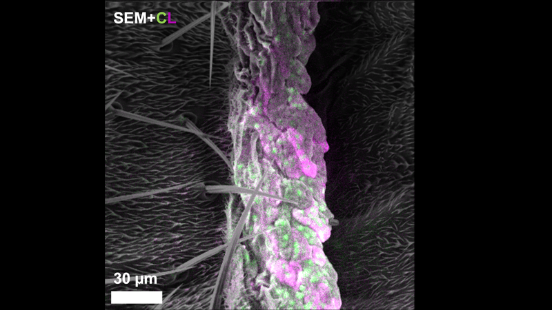
The team’s secret sauce? A superhero-like electron beam that triggers glowing probes attached to proteins. When zapped, these tags light up like tiny neon signs (thanks to cathodoluminescence—say that three times fast). Already tested on fruit flies fighting fungus, it’s basically giving researchers X-ray vision for biology. 🦸♂️
What’s Next?
While currently 2D-only, the crew plans to go full Avatar with 3D cellular maps using cryo-electron microscopy. Future med students might study cancer or Alzheimer’s with tech that makes today’s textbooks look like cave paintings. 📚→🎮